The
Paper Wasps and Hornets of Florida (Hymenoptera: Vespidae: Polistinae &
Vespinae) By Hugo Kons Jr. & Rex Rowan
APPENDIX C: Male Genitalia and Terminal
Metasomal Segments of Florida Polistes Species
Version 2018.1
Images of the voucher specimens for KOH treated dissections appear in Appendix D.
The dissection images include two non Florida species shown for
comparative purposes: Polistes instabilis
and Polistes aurifer. Also there are some non Florida specimens
of species that occur in Florida, as shown in Appendix D.
Click on the thumbnails to view individual figures, or scroll down for
the full set of figures sequentially with captions.
All Figures (C1-60) in order
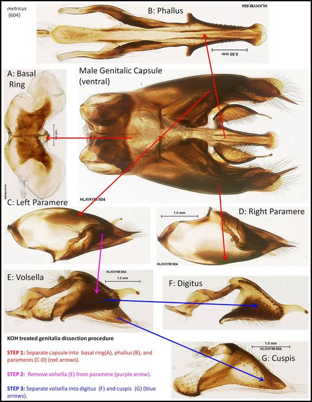
Figure C1: Components from dissecting apart the male genitalia capsule of a Polistes specimen after treatment with KOH.
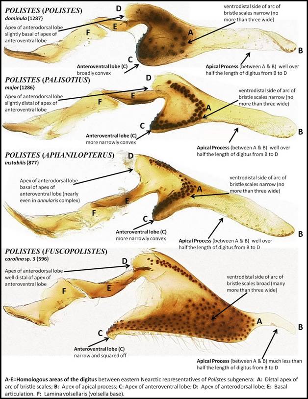
Figure C2: Comparative morphology of the digitus between Polistes subgenera occurring in the eastern Nearctic.
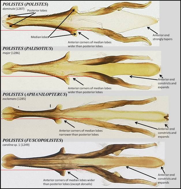
Figure C3: Comparative morphology of the phallus between Polistes subgenera occurring in the eastern Nearctic.
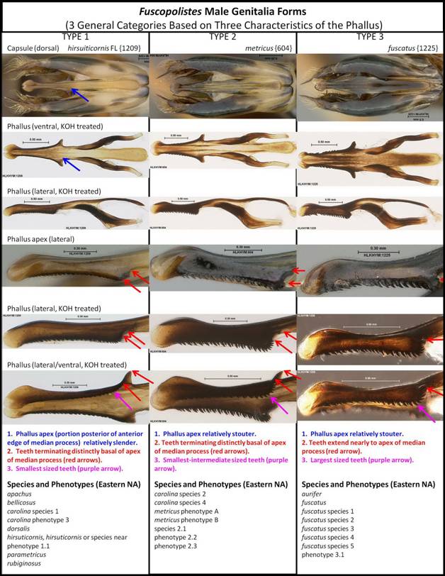
Figure C4: Three types of male genitalia that occur in Fuscopolistes, based on three characters of the phallus.
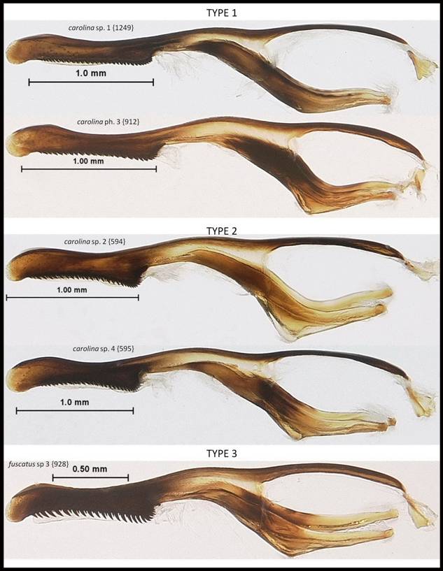
Figure C5: Examples of each of the three Fuscopolistes phallus types in lateral aspect.
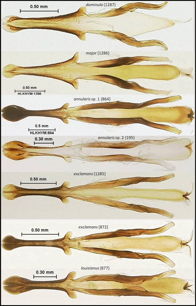
Figure C6: Phallus (ventral aspect) of Florida Polistes species (part 1 of 4).
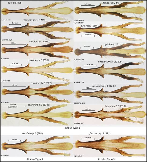
Figure C7: Phallus (ventral aspect) of Florida Polistes species (part 2 of 4).
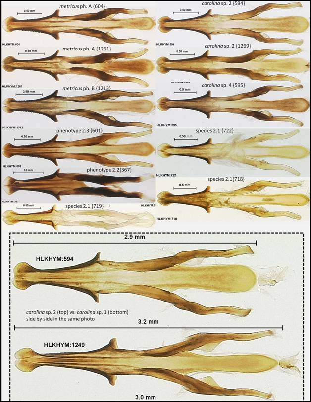
Figure C8: Phallus (ventral aspect) of Florida Polistes species (part 3 of 4).
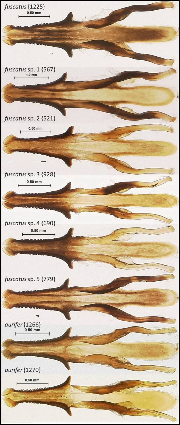
Figure C9: Phallus (ventral aspect) of Florida Polistes species (part 4 of 4).
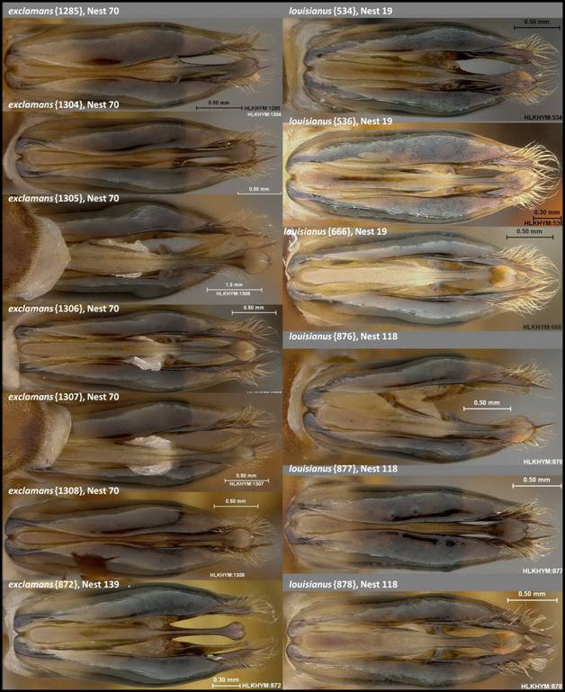
Figure C10: Comparison of genitalic capsule (dorsal aspect) between Polistes exclamans and Polistes louisianus, showing the posterior portion of the phallus. Note the phallus is not perfectly flat in these photos so the length and shape do may appear somewhat distorted (versus Figure C6 where the phallus in ventral aspect is flat against the bottom of a petri dish and held in place by a convex piece of glass).
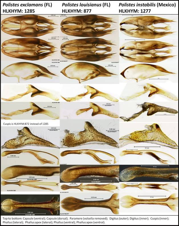
Figure C11: Comparison of male genitalic features between Polistes exclamans, P. louisianus, and P. instabilis (from Mexico). Despite the similarity in pattern between allopatric P. louisianus and P. instabilis, the phallus of P. instabilis is most like P. exclamans. Other features are similar (probably indistinguishable) between all three species.
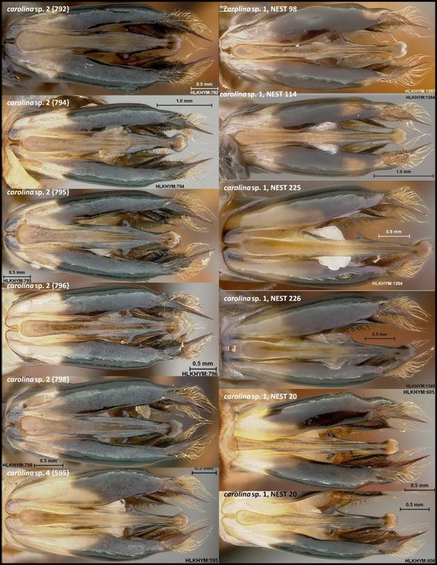
Figure C12: Comparison of genitalic capsule (dorsal aspect) between Polistes carolina sp. 1 and Polistes carolina sp. 2, showing the consistently stouter apical portion of the phallus in species 2. One P. carolina sp. 4 is also shown for comparison (see Figure C8 for comparisons of the phallus between these three species from KOH treated and dissected specimens).
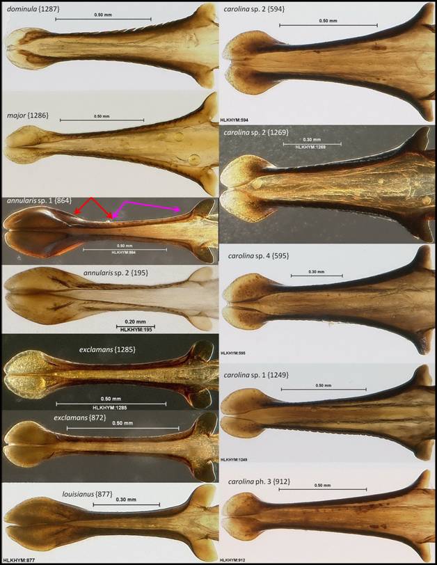
Figure C13: Phallus apex (ventral aspect) of Florida Polistes species (part 1 of 4).
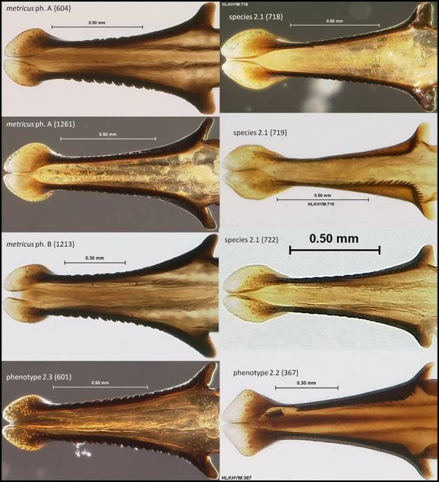
Figure C14: Phallus apex (ventral aspect) of Florida Polistes species (part 2 of 4).
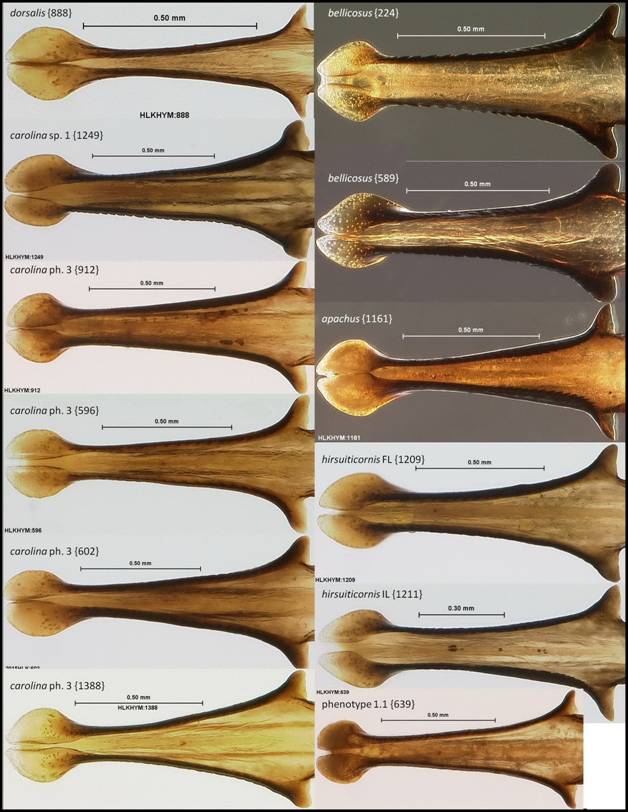
Figure C15: Phallus apex (ventral aspect) of Florida Polistes species (part 3 of 4).
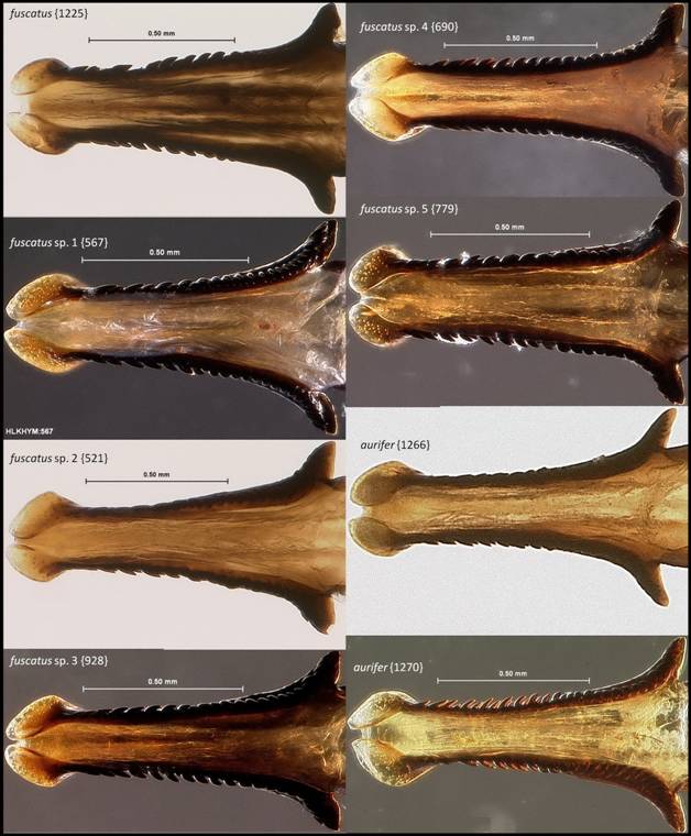
Figure C16: Phallus apex (ventral aspect) of Florida Polistes species (part 4 of 4).
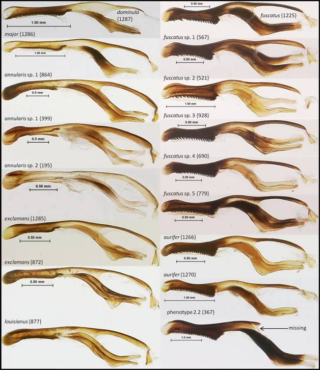
Figure C17: Phallus (lateral aspect) of Florida Polistes species (part 1 of 2).
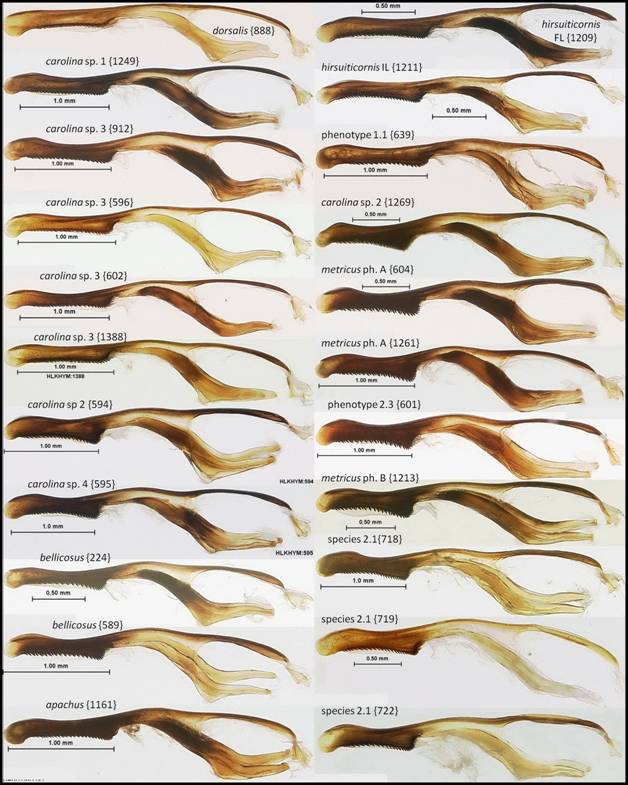
Figure C18: Phallus (lateral aspect) of Florida Polistes species (part 2 of 2).
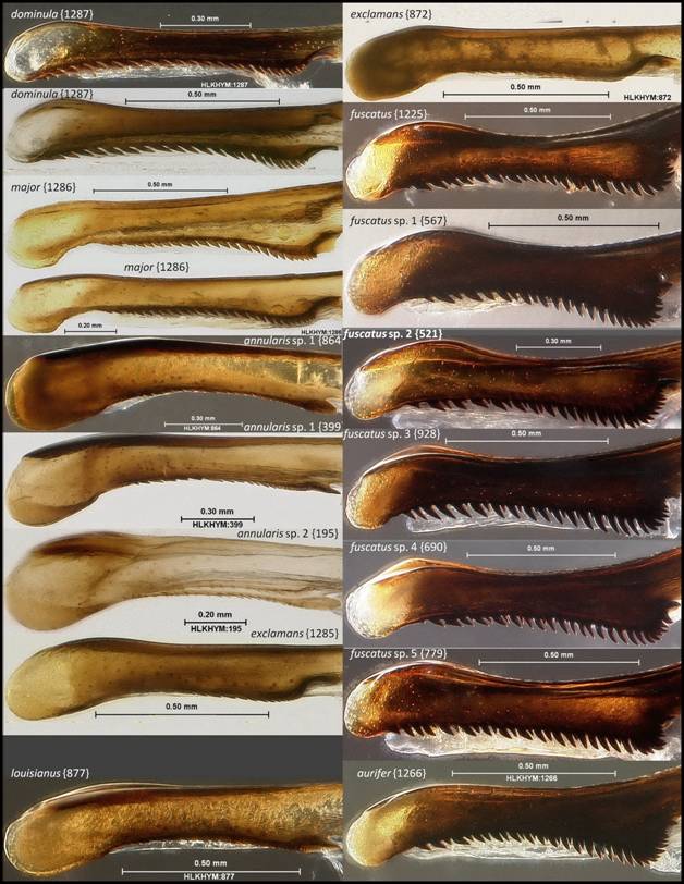
Figure C19: Phallus apex (lateral aspect) of Florida Polistes species (part 1 of 3).
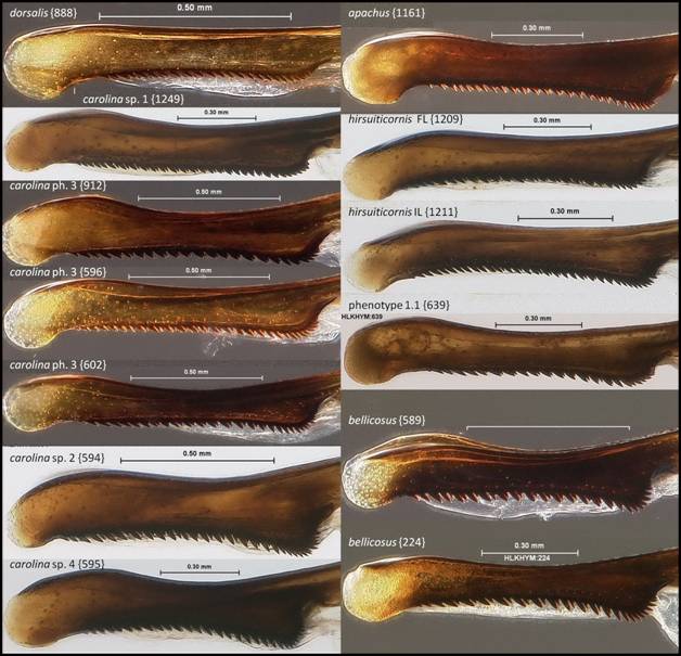
Figure C20: Phallus apex (lateral aspect) of Florida Polistes species (part 2 of 3).
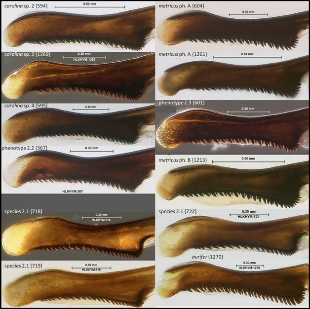
Figure C21: Phallus apex (lateral aspect) of Florida Polistes species (part 3 of 3).
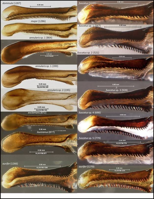
Figure C22: Phallus apex (tilted lateral aspect) of Florida Polistes species (part 1 of 3).
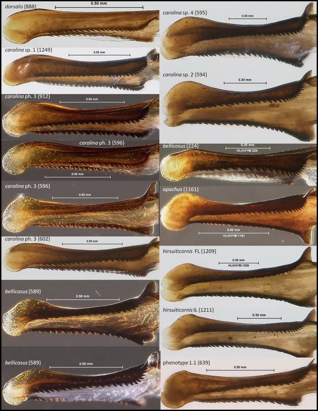
Figure C23: Phallus apex (tilted lateral aspect) of Florida Polistes species (part 2 of 3).
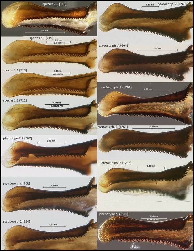
Figure C24: Phallus apex (tilted lateral aspect) of Florida Polistes species (part 3 of 3).
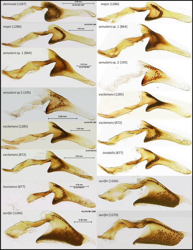
Figure C25: Digitus (inner side (on left), outer side (on right), lateral aspect) of Florida Polistes species (part 1 of 3 for inner and outer sides).
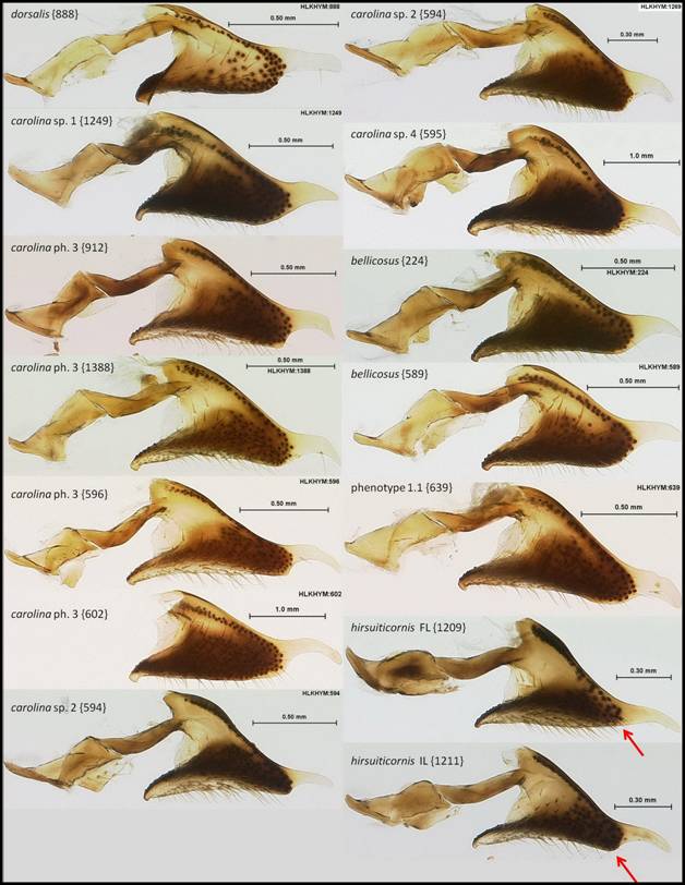
Figure C26: Digitus (inner side, lateral aspect) of Florida Polistes species (part 2 of 3 for inner side).
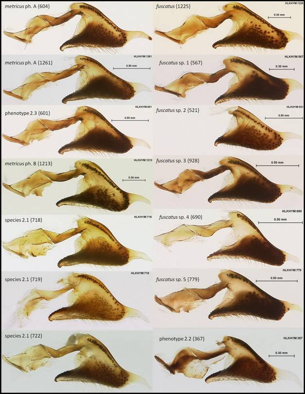
Figure C27: Digitus (inner side, lateral aspect) of Florida Polistes species (part 3 of 3 for inner side).
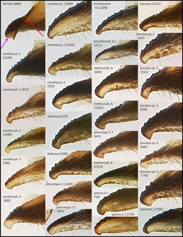
Figure C28: Antereoventral lobe of the digitus (inner side) of Florida Fuscopolistes species.
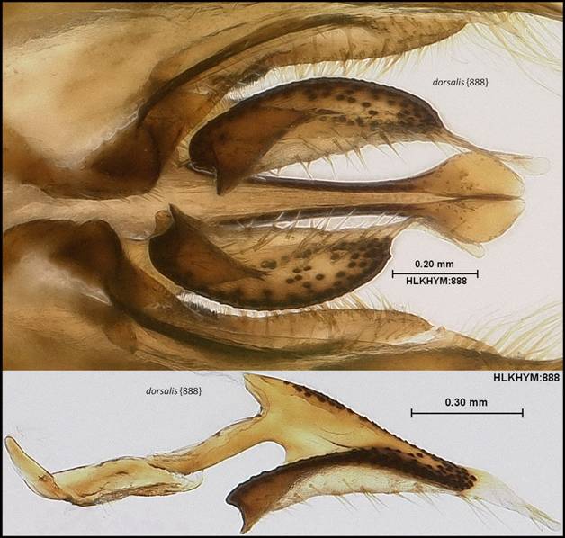
Figure C29: Polistes dorsalis. TOP: Each digitus in ventral aspect, showing concave antereoventral lobe and carina (on inner side of anterior portion of each digitus). BOTTOM: The outer side of a digitus (tilted lateral aspect) showing concave antereoventral lobe.
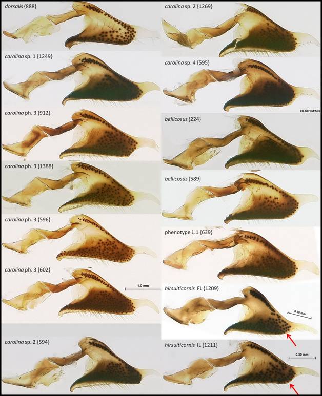
Figure C30: Digitus (outer side, lateral aspect) of Florida Polistes species (part 2 of 3 for outer side).
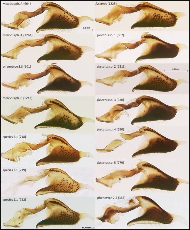
Figure C31: Digitus (outer side, lateral aspect) of Florida Polistes species (part 3 of 3 for outer side).
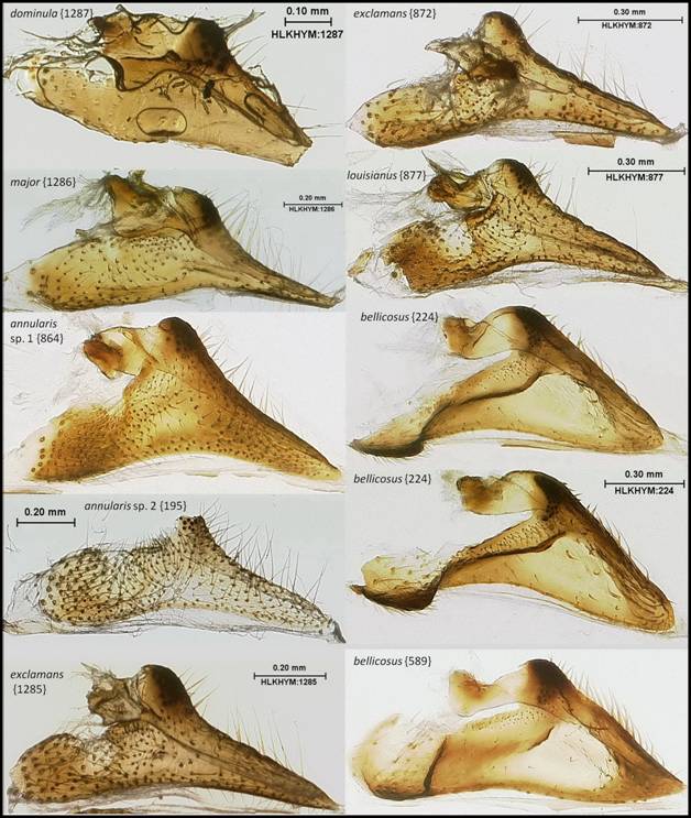
Figure C32: Cuspis (outer side, lateral aspect) of Florida Polistes species (part 1 of 4).
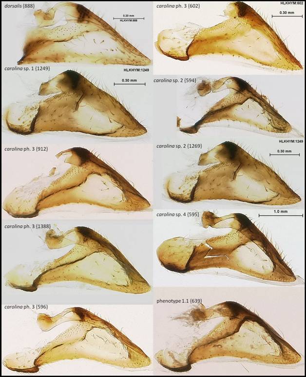
Figure C33: Cuspis (outer side, lateral aspect) of Florida Polistes species (part 2 of 4).
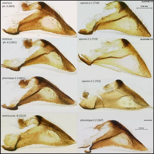
Figure C34: Cuspis (outer side, lateral aspect) of Florida Polistes species (part 3 of 4).
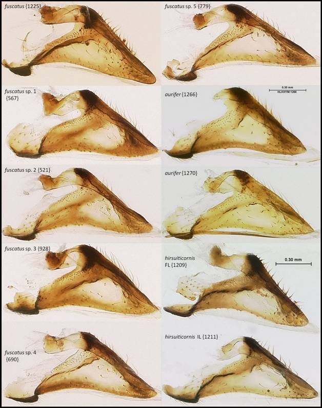
Figure
C35: Cuspis (outer side, lateral
aspect) of Florida Polistes species
(part 4 of 4).
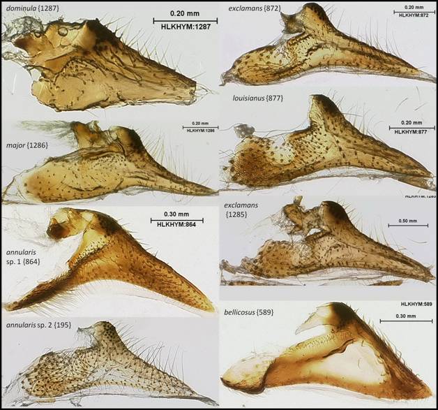
Figure
C36: Cuspis (inner side, lateral
aspect) of Florida Polistes species
(part 1 of 4).
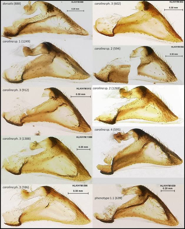
Figure
C37: Cuspis (inner side, lateral
aspect) of Florida Polistes species
(part 1 of 4).
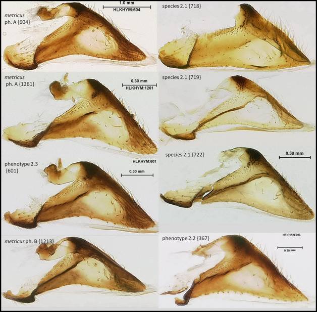
Figure
C38: Cuspis (inner side, lateral
aspect) of Florida Polistes species
(part 1 of 4).
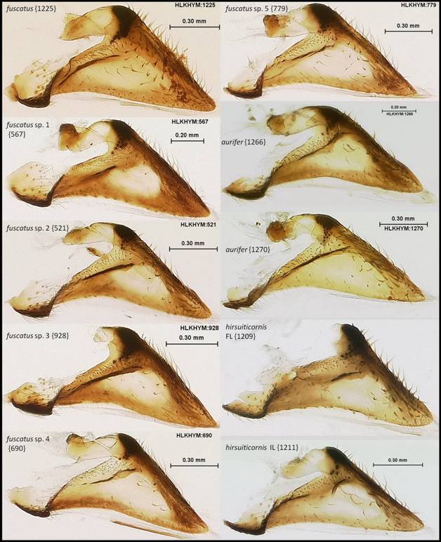
Figure
C39: Cuspis (inner side, lateral
aspect) of Florida Polistes species
(part 1 of 4).
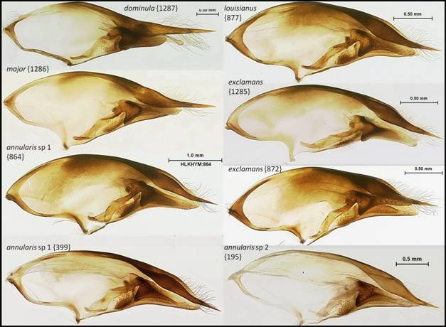
Figure C40: Paramere with volsella (inner side, lateral aspect) of Florida Polistes species (part 1 of 4).
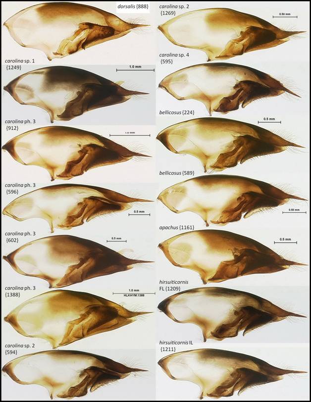
Figure C41: Paramere with volsella (inner side, lateral aspect) of Florida Polistes species (part 2 of 4).
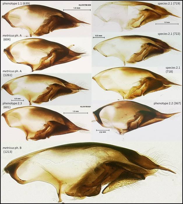
Figure C42: Paramere with volsella (inner side, lateral aspect) of Florida Polistes species (part 3 of 4).
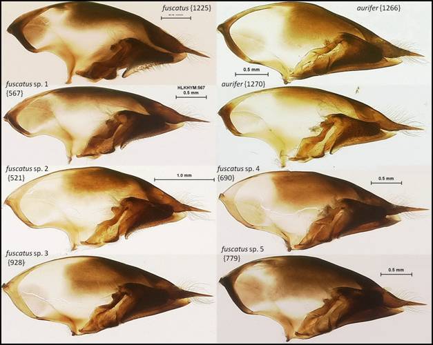
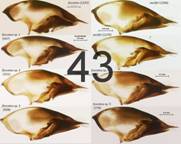
Figure C43: Paramere with volsella (inner side, lateral aspect) of Florida Polistes species (part 4 of 4).
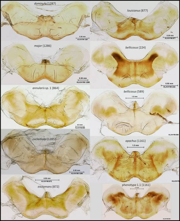
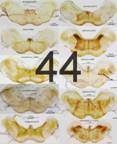
Figure C44: Basal ring (=gonocardo) (flattened on slide) of Florida Polistes species (part 1 of 4).
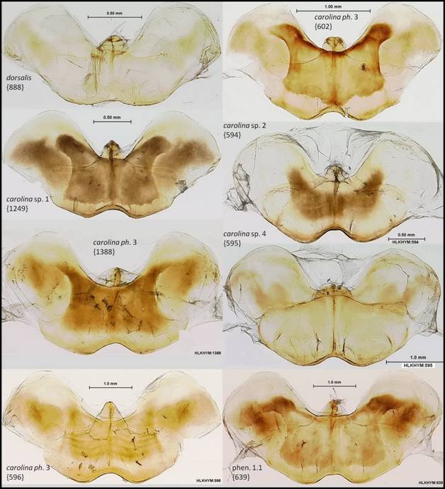
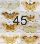
Figure C45: Basal ring (=gonocardo) (flattened on slide) of Florida Polistes species (part 2 of 4).
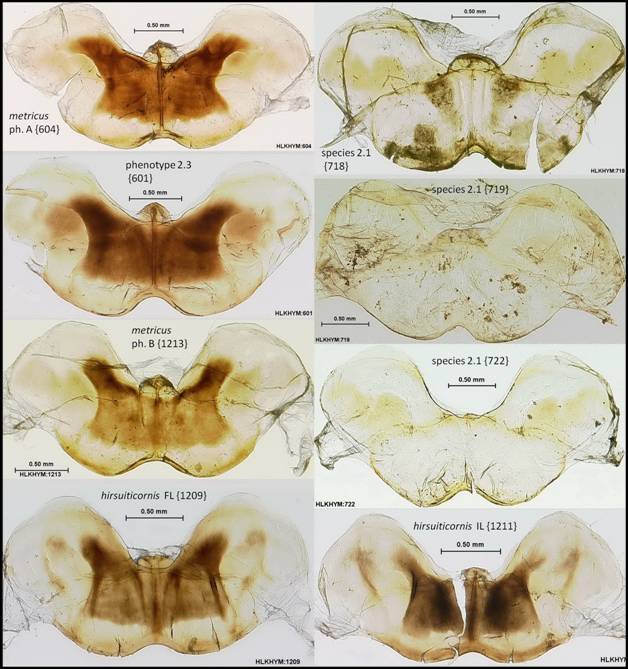
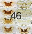
Figure C46: Basal ring (=gonocardo) (flattened on slide) of Florida Polistes species (part 3 of 4).
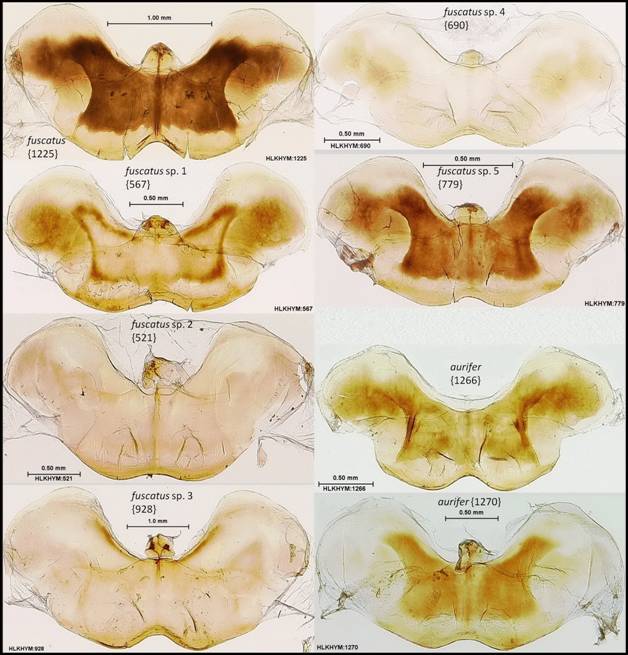
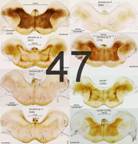
Figure C47: Basal ring (=gonocardo) (flattened on slide) of Florida Polistes species (part 4 of 4).
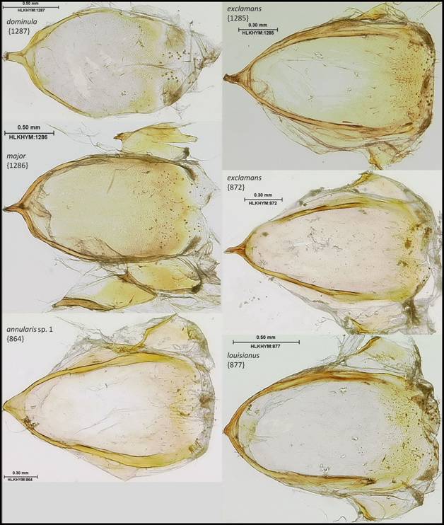
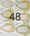
Figure C48: Segment X (flattened on slide) of Florida Polistes species (part 1 of 5).
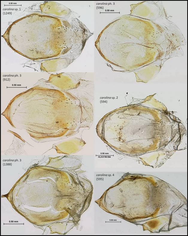
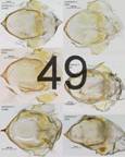
Figure C49: Segment X (flattened on slide) of Florida Polistes species (part 2 of 5).
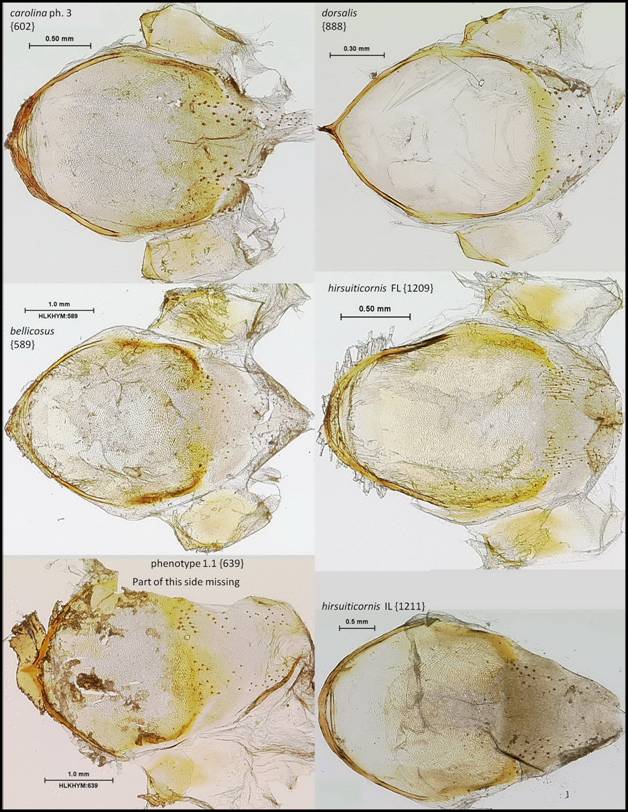
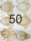
Figure C50: Segment X (flattened on slide) of Florida Polistes species (part 3 of 5).
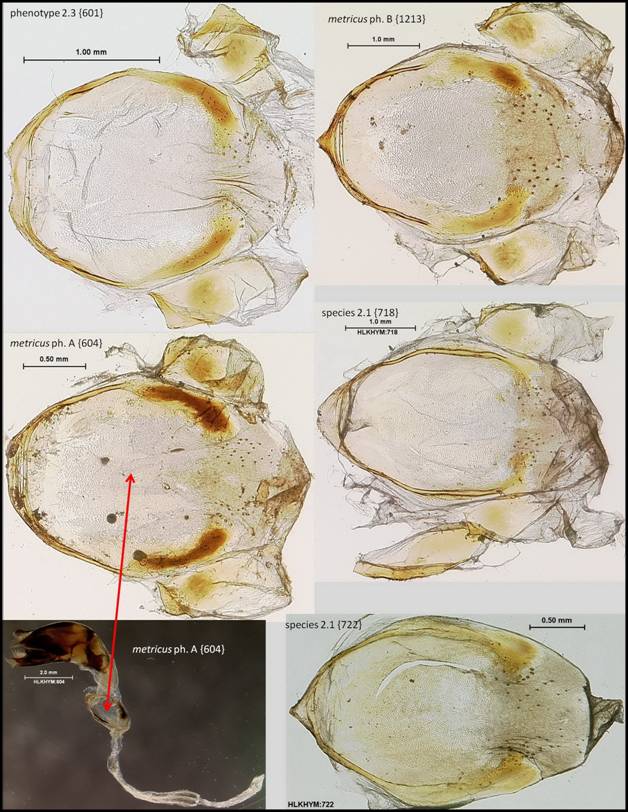
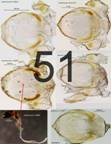
Figure C51: Segment X (flattened on slide) of Florida Polistes species (part 4 of 5).
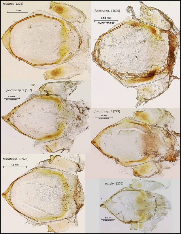
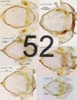
Figure C52: Segment X (flattened on slide) of Florida Polistes species (part 5 of 5).
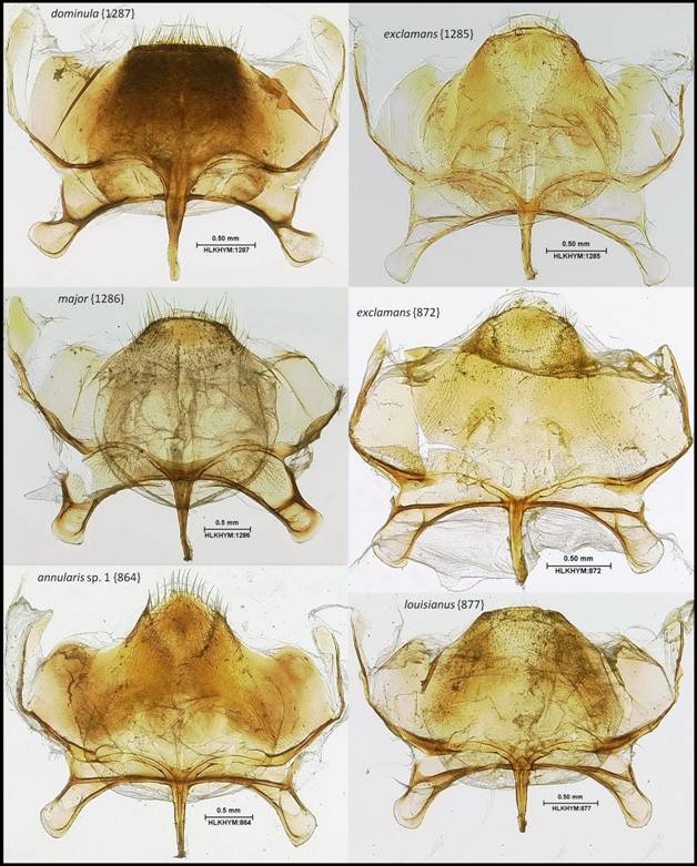
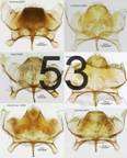
Figure C53: Sternite VIII+IX (KOH treated, flattened on slide, with lateral tergites IX connected on at least one side (except in 872)) of Florida Polistes species (part 1 of 2).
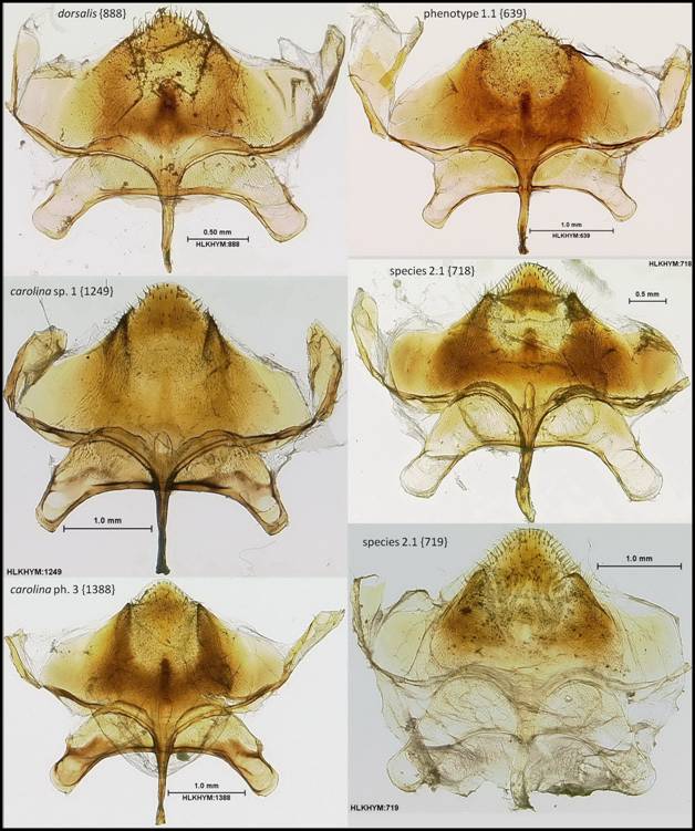
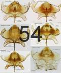
Figure C54: Sternite VIII+IX (KOH treated, flattened on slide, with lateral tergites IX connected on at least one side) of Florida Polistes species (part 2 of 2).
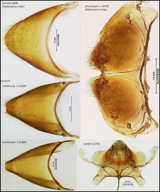
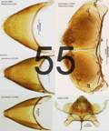
Figure C55: Tergite VIII (left column, right column top and middle) and sternite VIII+IX (right column, bottom) of Florida Polistes species. All are KOH treated, carolina complex images are not flattened on slide (column 1, middle and bottom [1249 & 1388]) whereas the other three are [888, 639, & 1270].
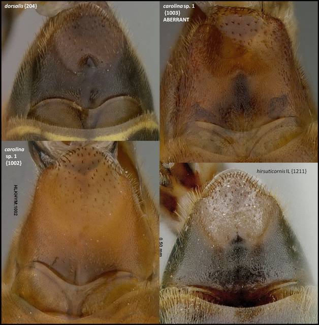
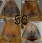
Figure
C56: Sternite VIII+IX (partially extracted from metasoma and not KOH
treated) of Florida Polistes species
(part 1 of 5).
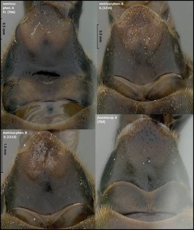
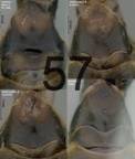
Figure
C57: Sternite VIII+IX (partially extracted from metasoma and not KOH
treated) of Florida Polistes species
(part 2 of 5).
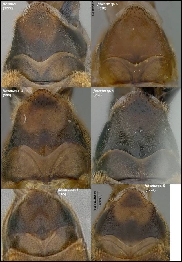
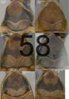
Figure
C58: Sternite VIII+IX (partially extracted from metasoma and not KOH
treated) of Florida Polistes species
(part 3 of 5).
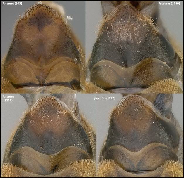
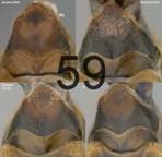
Figure
C59: Sternite VIII+IX (partially extracted from metasoma and not KOH
treated) of Florida Polistes species
(part 4 of 5).
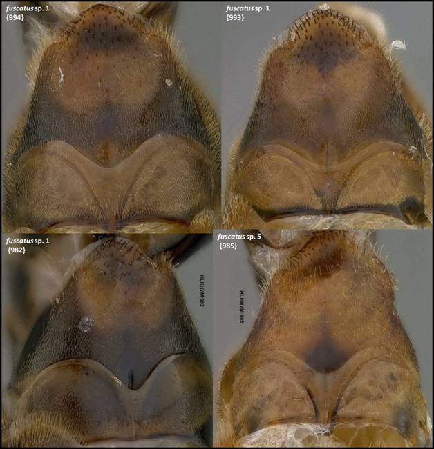
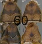
Figure
C60: Sternite VIII+IX (partially extracted from metasoma and not KOH
treated) of Florida Polistes species
(part 5 of 5).


